

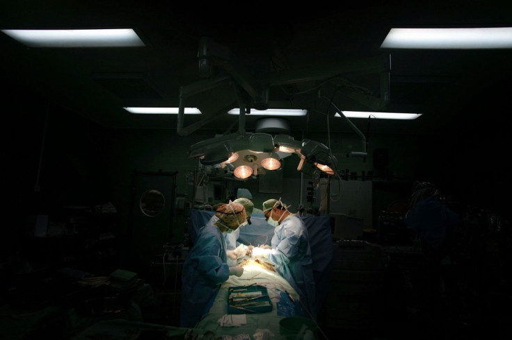
Surgeons can now use 3D printed hearts to practise
With the help of three-dimensional, or 3D printed models of the heart, surgeons will now able to physically assess patients’ hearts before opening them up for an operation. Researchers at the Massachusetts Institute of Technology (MIT) have successfully developed a new system that produces highly realistic, personalised 3D models of the blood-pumping organ, sourced from MRI scans.
The system, co-developed with researchers at the Boston Children’s Hospital, may address the worries of patients who will undergo heart surgery in the future, as well as their families. According to the team, it will make patients feel considerably safer knowing that that surgeons have already planned the operation ahead of time. With the new technique, a tangible model of the heart can be produced in just a few hours, enabling physicians to prepare for the anatomical idiosyncrasies of individual patients prior to a medical procedure.
Developers of this system used several different innovations, including software capable of independently analysing MRI scans, as well as new procedures to improve the precision of MRI scans tenfold. With the new method, human intervention is kept to a minimum, with algorithms left to infer the rest. This is a stark contrast to previous attempts at producing printable models, which required extensive manual involvement from human experts to determine the boundaries of individual MRI cross sections.
According to Polina Golland, a professor of electrical engineering and computer science at MIT and who is part of the team that developed this latest innovation, the team was surprised with the success of the new 3D method.
"I think that if somebody told me that I could segment the whole heart from eight slices out of 200, I would not have believed them. Our collaborators are convinced that this will make a difference. The phrase I heard is that ‘surgeons see with their hands,’ that the perception is in the touch," Golland says.
The new system will be presented at the International Conference on Medical Image Computing and Computer Assisted Intervention in Munich, Germany in October. A group of cardiac surgeons at the Boston Children’s Hospital will evaluate the technique using 3D models based on the hearts of 10 patients who have already received treatment. This is to determine whether the 3D models can improve surgical outcomes by comparing it with operations that were conducted in real-time without 3D mock-ups.
In October 2014, surgeons at Morgan Stanley Children’s Hospital in New York City saved the life of a two-week-old baby who required complicated heart surgery with the help of 3D printing. While surgery was deemed complicated and dangerous because the child's heart was riddled with holes and structured unusually, a 3D printed copy of the organ provided the surgeons the opportunity to study it and develop a detailed surgery strategy. This technique provided them with a road map to serve as a reliable guide, enabling the doctors to repair the baby’s heart with one operation.
Contact the writer at feedback@ibtimes.com.au or tell us what you think below.


