Researchers reveal new 3D structure of biologically active DNA more dynamic than 'Watson-Crick' double helix
A team of researchers at the Baylor College of Medicine in the US and University of Leeds in the UK have imaged a new three-dimensional structure of the supercoiled DNA using the microscopy technique.
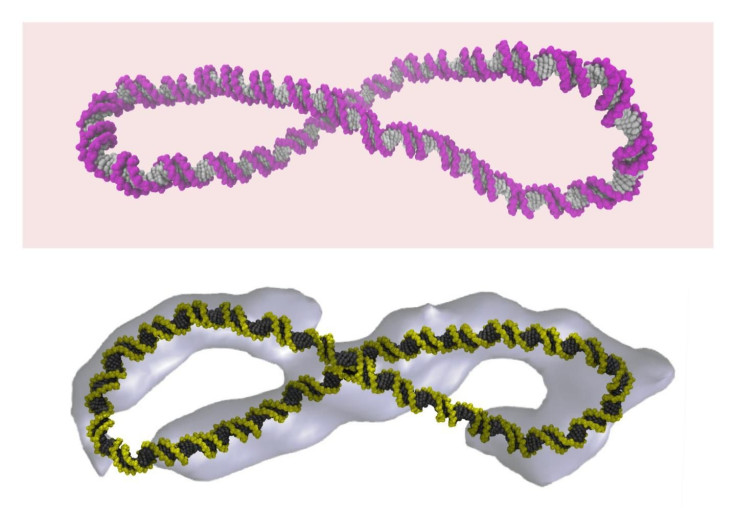
The new structure of the DNA has revealed that its shape is much more dynamic than what was historically explained by the famous “Watson-Crick” double helix model.
As described in the journal, Nature Communications, the 3D model shows the long strands of DNA are always in a constant wriggling motion. In addition, the strands form different shapes. The 3D model contradicts the double helix structure which suggests that the DNA is static and rigid.
The researchers used powerful microscopy technique to image the different shapes of the DNA. The team at the Baylor College of Medicine imaged the DNA, while researchers at the University of Leeds examined the image using supercomputer simulations at the campus.
"When Watson and Crick described the DNA double helix, they were looking at a tiny part of a real genome, only about one turn of the double helix,” said Dr Sarah Harris of the University of Leeds. "Our study looks at DNA on a somewhat grander scale - several hundreds of base pairs - and even this relatively modest increase in size reveals a whole new richness in the behaviour of the DNA molecule."
To study how DNA looked inside a cell, the researchers created circles of DNA so that its end trapped different degrees of winding. They then wound and unwound the tiny circles, making sure that they resembled the biologically active full-length DNA strands in the human cell.
Using the cryo-electron tomography, the researchers imaged the tiny circles and discovered that coiling led to the formation of different and unexpected shapes. The images of the different shapes were then subjected to computer simulation which explained that a dynamic motion leads to a constant changing of shape.
The researchers believe that the understanding of how DNA behaves inside a cell will help develop targeted therapies and better medicines and antibiotics for cancer treatment.
Contact the writer at feedback@ibtimes.com.au, or let us know what you think below.
- MOST POPULAR IN Health & Wellbeing