

Digital Frankenstein: Scientists slice female corpse 5,000 times for 'virtual human' project
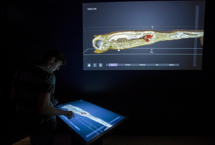
To conduct experiments too risky to try on living humans, scientists created a model by cutting a dead woman’s body into very thin slices and reconstructing it as the world’s most detailed digital body. The woman has been sliced more than 5,000 times, called the “human phantom,” for other researchers to conduct experiments without living human subjects.
The work is under the Visible Human Project, which is a long-running scientific effort to provide cross-sectional imagery of the human body. The American woman’s body was sliced just a third of a millimetre thick to be photographed in detail through MRI and CT scans.
The woman was believed to be obese who died from heart disease at the age of 59 in Maryland in the U.S, which were the only detail available about the corpse. Reports also show that her husband donated her body to the US National Library of Medicine in Bethesda, Maryland for the project.
Another body was also used for the Visible Human Project, which was sliced at cross-sectional thicknesses of 1mm. It was from a man named Joseph Paul Jernigan, who had been a convicted murderer and sentenced to death by lethal injection in Texas in 1993.
To date, the cross-sectional images of the bodies were compiled by a new research team. The images were enhanced into digitalised high-resolution imagery to create a “virtual human” for further study and experimental purposes on diseases and health conditions.
Ara Nazarian, researcher and an orthopaedic surgeon at Harvard Medical School, said that the images can give great opportunity to study human tissues without requiring studies on live humans, which are lengthy and expensive. The human phantom will now allow anyone to perform experiments on their laptop, she added.
In addition, Fernando Bello, a reader surgical graphics and computing expert at Imperial College London, told the New Scientist that researchers can also look for more information about organs and its structuring through the human phantom with 10 times as much information as they could get from an MRI scan. The research, presented at the annual conference of the IEEE Engineering in Medicine and Biology Society in Italy in August, indicated that aside from the safety benefits from the human phantom, the images could also provide massive benefits on experiment cost and practicality issues.
The virtual human would also allow doctors to develop safer, more effective scanning procedures for people with implants, the Science Alert reported. The team is currently using the images to analyse obesity and the impact of long-term cellphone use on the brain.
Lead researcher Sergey Makarov, of the Worcester Polytechnic Institute in the US, said that they are also hoping to improve breast cancer screening to provide better, more reliable mammogram results. The virtual human are freely available online. It could be modified though basic software used in labs around the world.
Contact the writer at feedback@ibtimes.com.au or tell us what you think below
-

Blinken Due In China Seeking Pressure But Also Stability
-

Israel Says US Military Aid Sends 'Strong Message' To Enemies
-
Chinese Sellers Go To TikTok School To Reach Buyers Abroad
-

Long-delayed Ukraine Aid Clears US Congress, Awaits Biden Signature
-

They Stormed The US Capitol In 2021 - Now They Want To Serve There


